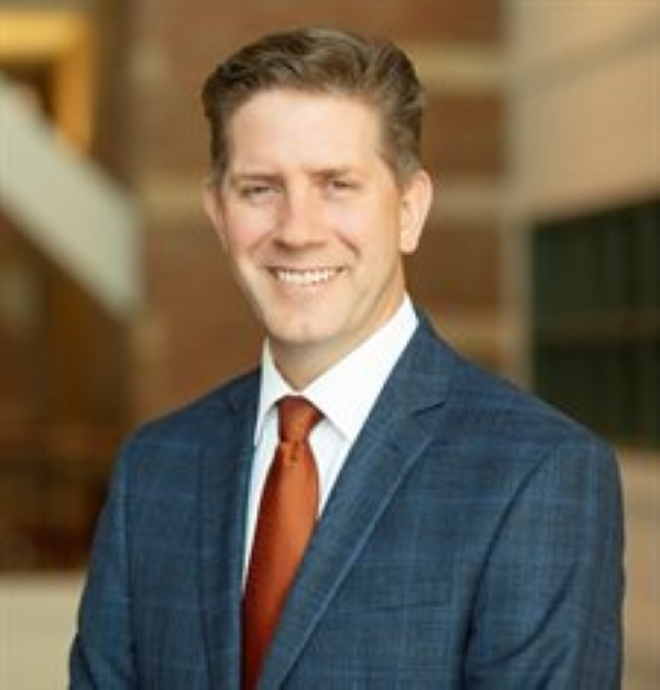
Biological Physics (iPoLS) Seminar: Stephen Boppart
- Event Type
- Seminar/Symposium
- Sponsor
- Center for Biophysics and Quantitative Biology
- Location
- Beckman Institute Room 3269 (3rd Floor Tower Room)
- Date
- Oct 21, 2022 2:00 pm
- Speaker
- Stephen Boppart, Professor, Grainger Distinguished Chair of Engineering, Director, GSK Center for Optical Molecular Imaging, Interim Director, Interdisciplinary Health Sciences Institute, University of Illinois Urbana Champaign.
- Contact
- Sharlene Denos
- denos@illinois.edu
- Views
- 26
- Originating Calendar
- Beckman Institute Calendar (internal events only)
Title: “Label-Free Multimodal Multiphoton Imaging for Probing Microscopic Dynamics in Living Systems.”
Speaker: Stephen Boppart, Professor, Grainger Distinguished Chair of Engineering, Director, GSK Center for Optical Molecular Imaging, Interim Director, Interdisciplinary Health Sciences Institute, University of Illinois Urbana Champaign.
Innovations in biomedical imaging have historically led to discoveries in the life sciences and new detection and diagnostic technologies in medicine and surgery. Label-free intravital optical imaging, in vitro imaging of cells and multi-cellular constructs, and imaging of fresh, unstained, resected tissue specimens, offer a wealth of new biosignatures for revealing biological and physiological, mechanisms, dynamics, and processes. Using innovative optical source technology and nonlinear optics to generate new excitation wavelengths and manipulate the light stimulus in new ways, Simultaneous Label-free Auto-fluorescence Multi-harmonic (SLAM) microscopy, Fluorescence Lifetime Imaging Microscopy (FLIM), and coherent Raman scattering (CRS) microscopy, can achieve fast simultaneous visualization of the rich intrinsic molecular and metabolic features within cells and tissues. Quantitative machine/deep learning analyses of these multi-dimensional datasets can be used to understand fundamental biological mechanisms as well as to identify selective biomarkers for pathology and disease states, such as cancer. Specifically, label-free hyperspectral coherent anti-Stokes Raman scattering (HS-CARS) microscopy was used to elucidate real-time dynamics of sub-cellular lipid droplets and mitochondria under various microenvironmental stressors and conditions, and tumor-associated extracellular vesicles (EVs) were analyzed via their optical signatures and spatial distributions. Quantitative analysis of the EVs showed that those from the tumor microenvironment have unique optical signatures, in comparison to those from healthy human subjects. The use of this platform of label-free nonlinear imaging techniques offers comprehensive cell-to-clinic capabilities, and the clinical demonstration of these optical biomedical imaging technologies offers new paradigms for point-of-procedure diagnosis and guidance.